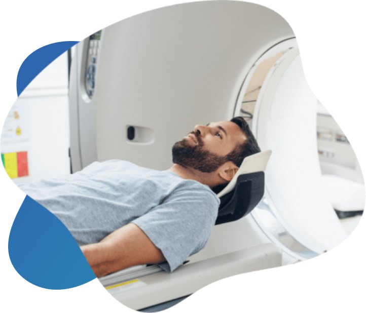
Medical professionals rely on advanced imaging techniques to evaluate patients after traumatic events. Internal injuries can present without visible external signs, making accurate assessment a priority in medical settings. Physicians use various tools to observe the body’s internal structures and determine the extent of trauma. CT scans offer a detailed view of these internal regions.
What Is a CT Scan?
A computed tomography scan, or a CT scan, represents a highly specialized form of X-ray technology. Standard radiographs provide two-dimensional pictures; these can work well for observing basic bone fractures. Computed tomography goes beyond this capability by capturing multidimensional views. The equipment produces cross-sectional images of the skeletal system, organs, muscles, and blood vessels.
These detailed visualizations allow medical teams to analyze complex structures and identify injuries within the human body. Because the machine captures both dense bone and soft tissue simultaneously, doctors receive a comprehensive overview of the internal anatomy. The resulting clarity helps physicians distinguish between overlapping structures that might blur together on a conventional X-ray.
How Does It Work?
The machinery features a large, circular opening designed to accommodate the patient, and individuals lie flat on a motorized examination table that glides smoothly through the center of this ring. As the table advances, an X-ray source rotates around the person, emitting narrow beams of radiation. Each rotation captures a single thin slice of the targeted anatomical region.
A computer system processes these individual cross-sectional images. By compiling many flat images, the software constructs comprehensive three-dimensional models. Radiology technologists review these detailed representations on their monitors. They can rotate the digital models to examine tissues from multiple perspectives and identify subtle anomalies hidden within the body’s crevices.
Some technologists administer a contrast material before the procedure, but the use of dye depends on the purpose of the imaging. This specialized dye highlights specific structures within the circulatory system or organs. As a result, blood vessels and targeted tissues stand out prominently against the surrounding anatomy.
What Can It Diagnose?
Medical providers order these tests to investigate a wide array of physical conditions and suspected traumas. Following an accident, the generated images may reveal signs of internal bleeding, organ damage, and complex bone fractures. The technology identifies severe head traumas; this can include skull damage and cerebral bleeding.
Beyond trauma evaluation, the scans assist in identifying chronic diseases and systemic disorders. Physicians rely on this detailed imaging to locate polyps, primary tumors, and metastatic cancers. The cross-sections may show indicators of gastrointestinal diseases, chronic sinusitis, and musculoskeletal disorders. Radiologists also look for arterial issues; these can present as aneurysms or blocked blood vessels. Respiratory conditions, like pneumonia and interstitial lung disease, may also appear on the resulting digital models. The capability to repeatedly monitor these conditions allows medical staff to track a patient’s response to ongoing treatments.
Seek Professional Radiology Services
Going to an imaging facility gives patients access to high-quality imaging and experienced medical personnel. Expert radiologists evaluate the final scans and send comprehensive reports directly to the referring physician. The referring doctor can then formulate a personalized treatment plan, basing it on these diagnostic findings.